Search results
Search for "X-ray absorption" in Full Text gives 66 result(s) in Beilstein Journal of Nanotechnology.
Properties of tin oxide films grown by atomic layer deposition from tin tetraiodide and ozone
Beilstein J. Nanotechnol. 2023, 14, 1085–1092, doi:10.3762/bjnano.14.89

- grazing incidence X-ray diffractometry (GIXRD), using an X-ray diffractometer SmartLab Rigaku with Cu Kα radiation, which corresponds to an X-ray wavelength of 0.15406 nm. X-ray photoelectron emission and X-ray absorption spectroscopy (XPS and XAS, respectively) measurements were made at the FinEstBeAMS
- eV binding energy, whereas the peak at 5 eV is not as dominant [28][29][35]. X-ray absorption spectra (Figure 11) were additionally recorded to possibly detect any differences between the samples. The Sn 3d XAS band is constituted by transitions from the 3d core orbital to the unoccupied p and f


Upscaling the urea method synthesis of CoAl layered double hydroxides
Beilstein J. Nanotechnol. 2023, 14, 927–938, doi:10.3762/bjnano.14.76

- process and aiming to quantify the amount of this Co-based α-LH impurity, X-ray absorption spectroscopy (XAS) measurements were performed at the CLÆSS BL22 beamline at the ALBA synchrotron. Figure 2A depicts the X-ray absorption near-edge structure (XANES) spectra for the Co K edge. In all samples, the
- of the structural features of the scale-up samples determined by extended X-ray absorption fine structure (EXAFS) measurements, the used model, and the corresponding fits can be found in Supporting Information File 1 (Figure S9 and Table S3). Thermal decomposition in both inert (nitrogen) and
- h at 523 K and 5 × 10⁻⁵ bar. The desorption branch of the N2 isotherm was used to determine the pore size distribution using the BJH method. The surface area was determined using the BET method. The micropores volumes were determined by applying t-plot and DR methods. X-ray absorption spectroscopy X


Evaluation of electrosynthesized reduced graphene oxide–Ni/Fe/Co-based (oxy)hydroxide catalysts towards the oxygen evolution reaction
Beilstein J. Nanotechnol. 2023, 14, 420–433, doi:10.3762/bjnano.14.34
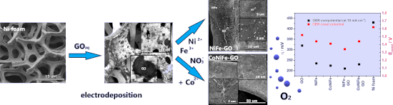
- probably inhibited the electrodeposition process of NiFe and CoNiFe on its surface. This may be the reason for the slower stabilization of the synthesis current density observed in the chronoamperograms (Figure 1a). X-ray diffraction, X-ray photoemission spectroscopy and X-ray absorption spectroscopy
- Figure 3a–d shows the X-ray absorption spectra (XAS) of the L3 edge of nickel (a), iron (b), cobalt (c), and carbon (d) in the studied catalysts. The appearance of a shoulder peak at the L3 edge of the nickel (Figure 3a) at 855 eV indicates the presence of oxides in the structure of the catalysts (Ni in
- ). Characterizations The morphology and structure of the catalysts were characterized using a scanning electron microscope (FEI QUANTA FEG 250) with an energy-dispersive X-ray (EDX) sensor. X-ray absorption spectroscopy (XAS) was performed at the 04BM beamline at the National Synchrotron Radiation Centre SOLARIS [41


Theoretical investigations of oxygen vacancy effects in nickel-doped zirconia from ab initio XANES spectroscopy at the oxygen K-edge
Beilstein J. Nanotechnol. 2022, 13, 975–985, doi:10.3762/bjnano.13.85

- Nanotechnologies, Institut National de Recherche en Sciences Exactes et Naturelles (IRSEN), Brazzaville, Congo 10.3762/bjnano.13.85 Abstract In this study, we present theoretical X-ray absorption near-edge structure (XANES) spectra at the K-edge of oxygen in zirconia containing Ni dopant atoms and O vacancies at
- and magnetic order in a typical diluted magnetic oxide. Such a finding may be crucial for spintronics-related applications. Keywords: defect; ligand field; nickel; oxidation state; oxides; spectroscopy; spintronics; vacancy; X-ray absorption; X-ray absorption near-edge structure (XANES); zirconia
- which is being currently employed in ultra-scaled electronics for its high dielectric constant [24][25] have received significant attention because of its practical applications. Thus, recently, exploiting first principles simulations and X-ray absorption near edge spectroscopy (XANES) in high magnetic


Revealing local structural properties of an atomically thin MoSe2 surface using optical microscopy
Beilstein J. Nanotechnol. 2022, 13, 572–581, doi:10.3762/bjnano.13.49

- deposited on a MoS2 crystal surface. Using near-edge X-ray absorption (NEXAFS), it was observed a strong dichroism in FePc thin films thicker than 4.5 nm. The strongest intensity of the N 1s→π* orbital transition at grazing incidence implies that the molecules are predominantly flat-lying with respect to


Investigation of electron-induced cross-linking of self-assembled monolayers by scanning tunneling microscopy
Beilstein J. Nanotechnol. 2022, 13, 462–471, doi:10.3762/bjnano.13.39

- separation [36][37][38][39][40][41][42]. The interactions between aromatic SAMs and electrons have been studied by X-ray photoelectron spectroscopy (XPS), near-edge X-ray-absorption fine-structure (NEXAFS) technique [43][44][45][46][47][48][49], infrared spectroscopy [43][50][51][52], high-resolution


Sputtering onto liquids: a critical review
Beilstein J. Nanotechnol. 2022, 13, 10–53, doi:10.3762/bjnano.13.2


A review on the green and sustainable synthesis of silver nanoparticles and one-dimensional silver nanostructures
Beilstein J. Nanotechnol. 2021, 12, 102–136, doi:10.3762/bjnano.12.9


Scanning tunneling microscopy and spectroscopy of rubrene on clean and graphene-covered metal surfaces
Beilstein J. Nanotechnol. 2020, 11, 1157–1167, doi:10.3762/bjnano.11.100

- near-edge X-ray absorption fine structure spectroscopy [15]. The observed chirality, R-C42H28 and L-C42H28, therefore results from the left or right pair of phenyl groups of a twisted C42H28 molecule being closer to the surface. On Au(111) the formation of homochiral clusters led to remarkable


Band tail state related photoluminescence and photoresponse of ZnMgO solid solution nanostructured films
Beilstein J. Nanotechnol. 2020, 11, 899–910, doi:10.3762/bjnano.11.75

- unavoidable in the structure transformation process, in a certain interval of Mg concentrations. The phase segregation process was investigated in detail by means of X-ray diffraction, element-specific near-edge X-ray absorption fine structure (NEXAFS), electron dispersive spectroscopy (EDS), atomic force


Hexagonal boron nitride: a review of the emerging material platform for single-photon sources and the spin–photon interface
Beilstein J. Nanotechnol. 2020, 11, 740–769, doi:10.3762/bjnano.11.61

- means, such as optical reflectance and absorption, electron energy loss spectroscopy, X-ray absorption, emission, and inelastic scattering. Regarding luminescence studies, PL is the light emission, i.e., the electromagnetic radiation from matter after the absorption of photons. It is originated by


Comparison of fresh and aged lithium iron phosphate cathodes using a tailored electrochemical strain microscopy technique
Beilstein J. Nanotechnol. 2020, 11, 583–596, doi:10.3762/bjnano.11.46

- (FIB) SEM and X-ray absorption near edge structure (XANES) [6][7][8][9][10][11][12][13]. Another technique for post-mortem analysis is atomic force microscopy (AFM). In its basic form, it provides information on the topography of the sample. More advanced AFM modes extract in addition to the topography


Mobility of charge carriers in self-assembled monolayers
Beilstein J. Nanotechnol. 2019, 10, 2449–2458, doi:10.3762/bjnano.10.235

- characterized immediately after the SAM preparation. For the Near-edge X-ray absorption fine structure (NEXAFS) measurements, gold-coated silicon wafer substrates were used. Gold films of 100 nm thickness were evaporated thermally at 453 K under high-vacuum conditions (≈10−7 mbar) with 5 nm titanium as the
- should be oriented almost perpendicular to the substrate surface and the phenyl groups are tilted to the substrate surface. The quantitative analysis of near-edge X-ray absorption fine structure (NEXAFS) data (Supporting Information File 1, Figure S4) reveals that the anthracene units within the SAMs


Green and scalable synthesis of nanocrystalline kuramite
Beilstein J. Nanotechnol. 2019, 10, 2073–2083, doi:10.3762/bjnano.10.202

- of nanocrystalline kuramite by means of a simpler, greener and scalable solvothermal synthesis. We exploited a multianalytical characterization approach (X-ray diffraction, extended X-ray absorption fine structure, field emission scanning electron microscopy, Raman spectroscopy and electronic
- microscopy (SEM), principal component analysis (PCA) of the wavelength dispersion spectroscopy (WDS) data, X-ray absorption spectroscopy (XAS) and Raman spectroscopy. Materials and Methods Synthesis The reactants necessary for the three syntheses are: CuCl2·2H2O (Merck), ZnCl2 (Merck), SnCl2·2H2O (Riedel-de
- ) were carried out at the LISA CRG beamline (BM-08; [57]) at the European Synchrotron Radiation Facility (ESRF, Grenoble, France). The software ATHENA [58] was used to average multiple spectra. Standard procedures [59] were followed to extract the structural extended X-ray absorption fine structure


The influence of porosity on nanoparticle formation in hierarchical aluminophosphates
Beilstein J. Nanotechnol. 2019, 10, 1952–1957, doi:10.3762/bjnano.10.191

- observed (Figure 3 and Figures S10–S15, Supporting Information File 1). There was good agreement with the Au foil, suggesting the gold has been successfully reduced to metallic gold particles. The Au/MP-SAPO-5 systems show a lower-energy X-ray absorption near edge structure (XANES), suggesting a higher


Materials nanoarchitectonics at two-dimensional liquid interfaces
Beilstein J. Nanotechnol. 2019, 10, 1559–1587, doi:10.3762/bjnano.10.153

- water phase, yielding a monolayer sheet of the two-dimensional nickel–iron cyanide grid network. Characterizations of the extended network by X-ray photoelectron spectroscopy (XPS), FTIR spectroscopy, SQUID magnetometry, X-ray absorption fine structure (XAFS), and grazing incidence synchrotron X-ray


Gas sensing properties of individual SnO2 nanowires and SnO2 sol–gel nanocomposites
Beilstein J. Nanotechnol. 2019, 10, 1380–1390, doi:10.3762/bjnano.10.136

- ; gas transport method; nanowires; quasi-one-dimensional materials; sol–gel synthesis; tin dioxide; X-ray absorption near edge structure (XANES); X-ray photoelectron spectroscopy (XPS); Introduction Semiconductor sensor functionality relies on heterogeneous catalytic chemical processes, which makes the
- calcination as follows: Figure 2 shows a TEM image of the obtained material. The particle diameter derived from this measurement was found to be 4–6 nm. X-ray spectroscopy of the materials In the present study, we used the non-destructive techniques, X-ray photoelectron spectroscopy (XPS) and X-ray absorption


Integration of LaMnO3+δ films on platinized silicon substrates for resistive switching applications by PI-MOCVD
Beilstein J. Nanotechnol. 2019, 10, 389–398, doi:10.3762/bjnano.10.38

- nanostructure growth was further analyzed in cross section by transmission electron microscopy (TEM), a JEOL 2011 equipment operating at 200 kV with a 0.19 nm point-to-point resolution. X-ray absorption near-edge spectroscopy (XANES) spectra at the Mn K-edge of LMO thin films were collected at the ESRF ID12


Enhancement of X-ray emission from nanocolloidal gold suspensions under double-pulse excitation
Beilstein J. Nanotechnol. 2018, 9, 2609–2617, doi:10.3762/bjnano.9.242

- ablation [6]. In practical applications for X-ray diffraction [7] and X-ray absorption fine structure (XAFS) measurements [8][9], or further nonlinear X-ray processes, a high flux of X-ray pulses is indispensable. X-ray intensity enhancements can be expected through an effective increase of the laser


Metal-free catalysis based on nitrogen-doped carbon nanomaterials: a photoelectron spectroscopy point of view
Beilstein J. Nanotechnol. 2018, 9, 2015–2031, doi:10.3762/bjnano.9.191

- ]: Niwa et al. [105] described carbon nanostructure alloys (a precursor of graphene), Nagaiah and co-workers studied nitrogen-doped CNTs [106], and Parvez et al. used N-graphene [107]. In the first report, correlating X-ray absorption spectroscopy (XAS) and ORR activity, the study was focused on the
- graphitic N was higher. Using X-ray absorption spectroscopy, the authors were able to identify different nitrogen configurations: three characteristic peaks were found at 399.1, 400.1 and 401.5 eV and were assigned to pyridinic N, cyanide (triple C–N bond) and graphitic N, respectively. This was in
- nitrogen plasma and studied its interaction with molecular oxygen. They showed that the interaction results in oxygen dissociation and the formation of carbon–oxygen single bonds on graphene, as observed by X-ray absorption spectroscopy of the carbon K edge and the O 1s core level. After the exposure to


Mechanistic insights into plasmonic photocatalysts in utilizing visible light
Beilstein J. Nanotechnol. 2018, 9, 628–648, doi:10.3762/bjnano.9.59

- excitation and electron injection into the semiconductor are still unclear. The verification that plasmon-excited electrons in Au NPs possess sufficient energy to overcome the Schottky junction to be injected into TiO2 was confirmed using high-resolution X-ray absorption spectroscopy (HR-XAS) [105]. The
- adopted experimental setup is depicted in Figure 10. The significant spectral variations observed by X-ray absorption spectroscopy (XAS) and resonant inelastic X-ray scattering (RIXS) suggest that electrons injected from Au NPs upon LSPR excitation could survive longer and become trapped at a Ti site near
- [6], copyright 2013 IOP Publishing. High-resolution X-ray absorption spectroscopy (HR-XAS) experiment used to determine the changes in the Au LIII-edge induced by 100 mW continuous wave laser excitation of the localized surface plasmon at 532 nm. Reprinted with permission from [105], copyright 2013


One-step chemical vapor deposition synthesis and supercapacitor performance of nitrogen-doped porous carbon–carbon nanotube hybrids
Beilstein J. Nanotechnol. 2017, 8, 2669–2679, doi:10.3762/bjnano.8.267

- was studied by means of X-ray photoelectron spectroscopy (XPS) and near-edge X-ray absorption fine structure (NEXAFS) at the facility of the Russian-German beamline at the Berliner Elektronenspeicherring für Synchrotronstrahlung (BESSY II) station. XPS spectra were measured at an energy of
- near-edge X-ray absorption fine structure (NEXAFS) N K-edge total-electron yield (TEY) spectra (b) of CNx materials synthesized using Fe/Mo, Ni/Mo, and Co/Mo catalysts. Raman spectra of CNx materials synthesized using Fe/Mo, Co/Mo, and Ni/Mo catalysts (a). Relationship between the ratios of Raman peak


(Metallo)porphyrins for potential materials science applications
Beilstein J. Nanotechnol. 2017, 8, 1786–1800, doi:10.3762/bjnano.8.180

- layers. The templating effect of the first layer can extend up to tens of nanometers in phthalocyanine layers [61]. In subsequent aging studies and in combination with near edge X-ray absorption fine structure (NEXAFS) spectroscopy we could show that the values of α apparently increase by 4–7° on both


Effect of the fluorination technique on the surface-fluorination patterning of double-walled carbon nanotubes
Beilstein J. Nanotechnol. 2017, 8, 1688–1698, doi:10.3762/bjnano.8.169

- show here that the C–F bond strength is sensitive to surroundings in the addition pattern, which can be controlled through the fluorination method. The preferable fluorine distributions on the DWCNT surface are proposed from quantum-chemical modelling of the fluorine near-edge X-ray absorption fine
- nanotubes (DWCNTs) are fluorinated using (1) fluorine F2 at 200 °C, (2) gaseous BrF3 at room temperature, and (3) CF4 radio-frequency plasma functionalization. These have been comparatively studied using transmission electron microscopy and infrared, Raman, X-ray photoelectron, and near-edge X-ray
- absorption fine structure (NEXAFS) spectroscopy. A formation of covalent C–F bonds and a considerable reduction in the intensity of radial breathing modes from the outer shells of DWCNTs are observed for all samples. Differences in the electronic state of fluorine and the C–F vibrations for three kinds of


Charge transfer from and to manganese phthalocyanine: bulk materials and interfaces
Beilstein J. Nanotechnol. 2017, 8, 1601–1615, doi:10.3762/bjnano.8.160

- photoemission spectroscopy (IPES), electron energy-loss spectroscopy (EELS), spectroscopic ellipsometry and X-ray absorption spectroscopy (XAS). Here, we only briefly mention the kind of information that is provided by these methods, and we refer the reader to comprehensive literature for detailed information
- revealed by polarization dependent X-ray absorption spectroscopy (XAS) studies [127]. In Figure 12 we present corresponding N 1s absorption spectra for pure MnPc and two different film thicknesses of an F16CoPc overlayer, deposited on a gold single crystal. Different light polarizations with respect to the
- represents the changes of the work function. Panel (b) focusses on the energy region close to the Fermi level. N 1s excitation spectra as obtained using X-ray absorption spectroscopy (adapted from [127]). Corresponding data for (c) a 2.0 nm thick MnPc film on Au(100), (b) an additional 0.6 nm thick F16CoPc


